An Overview of Fat Necrosis: Causes & Treatment
Explore the basics of fat necrosis including causes, treatment options, and common symptoms.
Published on May 3, 2024. Reviewed by Tara Call Triplett, RN, WCC, CHFN
Clinical Overview
Fat necrosis is a benign inflammatory condition that occurs when adipose (fat) tissue becomes damaged and dies. While it is not cancerous, fat necrosis can mimic more serious conditions, particularly in the breast, and may cause concern for patients and clinicians alike. For wound care professionals, understanding what causes fat necrosis, how it presents, and how to manage it is essential for accurate assessment, patient education, and appropriate referrals.
What is fat necrosis?
Fat necrosis occurs when the blood supply to fatty tissue is disrupted, leading to adipocyte death and a localized inflammatory response. The body replaces the damaged fat cells with scar tissue, calcifications, or oil cysts. This process can result from trauma, surgery, radiation therapy, or injections.
Common causes include:
- Blunt force trauma
- Surgical procedures, including reconstructive or cosmetic surgery
- Radiation therapy
- Injections or fat grafting
- Poor tissue perfusion
When patients ask, “What causes fat necrosis?” the simplest explanation is that it results from injury to fat tissue that compromises circulation and triggers inflammation.
Where does fat necrosis typically occur?
Fat necrosis most commonly develops in areas with abundant adipose tissue.
| Location | Typical Appearance | Risk / Severity Level |
| Breast (fat necrosis breast) | Firm or irregular lump; may cause skin dimpling, retraction, bruising, or calcifications on mammogram; may resemble malignancy on exam or imaging | Moderate clinical concern due to overlap with breast cancer; requires imaging and sometimes biopsy to rule out malignancy |
| Buttocks (fat necrosis after a BBL) |
Firm nodules, contour irregularities, localized swelling or tenderness; possible oil cyst formation or drainage if superficial | Low to moderate severity; typically benign but may require intervention if painful, infected, or cosmetically significant |
| Abdomen |
Palpable subcutaneous mass following surgery or trauma; may feel firm or slightly tender; occasionally associated with surgical scars | Low severity; usually self-limiting unless complicated by infection or wound breakdown |
| Thighs |
Localized firm areas after trauma, injections, or fat grafting may present with mild discoloration or induration. | Low severity; primarily monitored unless symptoms worsen |
| Flanks |
Subcutaneous firmness or nodularity, often post-trauma or post-surgical |
Low severity; conservative management is typically sufficient |
Areas exposed to radiation therapy |
Firm, fibrotic tissue changes; possible skin thickening; may coexist with scar tissue. |
Variable severity; may complicate wound healing in patients with radiation-induced tissue damage |
| Pressure-prone areas (e.g., pannus folds) | Induration beneath intact or compromised skin; may be mistaken for an abscess or deep tissue injury. | Variable severity; important to differentiate from infection or pressure injury |
Fat necrosis of the breast
Fat necrosis of the breast often follows lumpectomy, mastectomy with reconstruction, breast biopsy, reduction surgery, or radiation therapy. It may also occur after direct trauma, such as a seatbelt injury in a motor vehicle accident.
In wound care and post-surgical settings, clinicians should be aware that breast fat necrosis can present months after the inciting event.
Fat necrosis after a BBL
Fat necrosis after a BBL (Brazilian butt lift) is a known complication of fat grafting procedures. During a BBL, autologous fat is harvested and injected into the buttocks. If some of the transferred fat does not establish an adequate blood supply, it may undergo necrosis. This can result in firm nodules, contour irregularities, localized pain, or inflammation.
Wound care nurses may encounter these patients during post-procedure follow-up, particularly if complications such as infection, wound breakdown, or drainage occur.
Fat necrosis vs breast cancer on an ultrasound
In superficial areas, the lesion may feel hard and fixed, raising concern for malignancy. In the breast, fat necrosis may cause palpable masses that closely resemble breast cancer on physical examination.
Imaging findings vary depending on the stage of the lesion. On mammography, fat necrosis may appear as calcifications or a spiculated mass. This overlap in appearance often prompts further diagnostic evaluation.
Distinguishing fat necrosis from breast cancer on an ultrasound can be challenging. Fat necrosis may appear as:
- Complex cystic lesions
- Hypoechoic masses
- Areas with posterior acoustic shadowing
- Lesions with internal echogenic bands
Because these findings can overlap with malignancy, radiologists often correlate ultrasound with mammography and clinical history. A biopsy may be required if imaging is inconclusive. Wound care clinicians should reinforce the importance of follow-up and reassure patients that additional testing is precautionary.
Does fat necrosis go away?
A common question from patients is, “Does fat necrosis go away?”
In many cases, yes. Fat necrosis is benign and may resolve spontaneously over time. The body gradually reabsorbs damaged fat, and the lump may shrink or soften over months to years. However, some areas may persist as firm scar tissue or calcifications.
Resolution depends on:
- Size of the lesion
- Extent of tissue damage
- Overall vascular supply
- Presence of secondary complications
If the mass is stable, asymptomatic, and imaging confirms a benign diagnosis, conservative management is typically appropriate.
What should a wound care nurse know?
Wound care nurses play an important role in recognizing fat necrosis, particularly in postoperative and trauma patients. Key considerations include:
Thorough history-taking
Document recent surgeries, radiation therapy, trauma, injections, or cosmetic procedures such as fat grafting.
Assessment of skin integrity
Evaluate for erythema, fluctuance, drainage, warmth, or signs of infection. Although fat necrosis itself is not infectious, secondary infection can occur.
Monitoring for complications
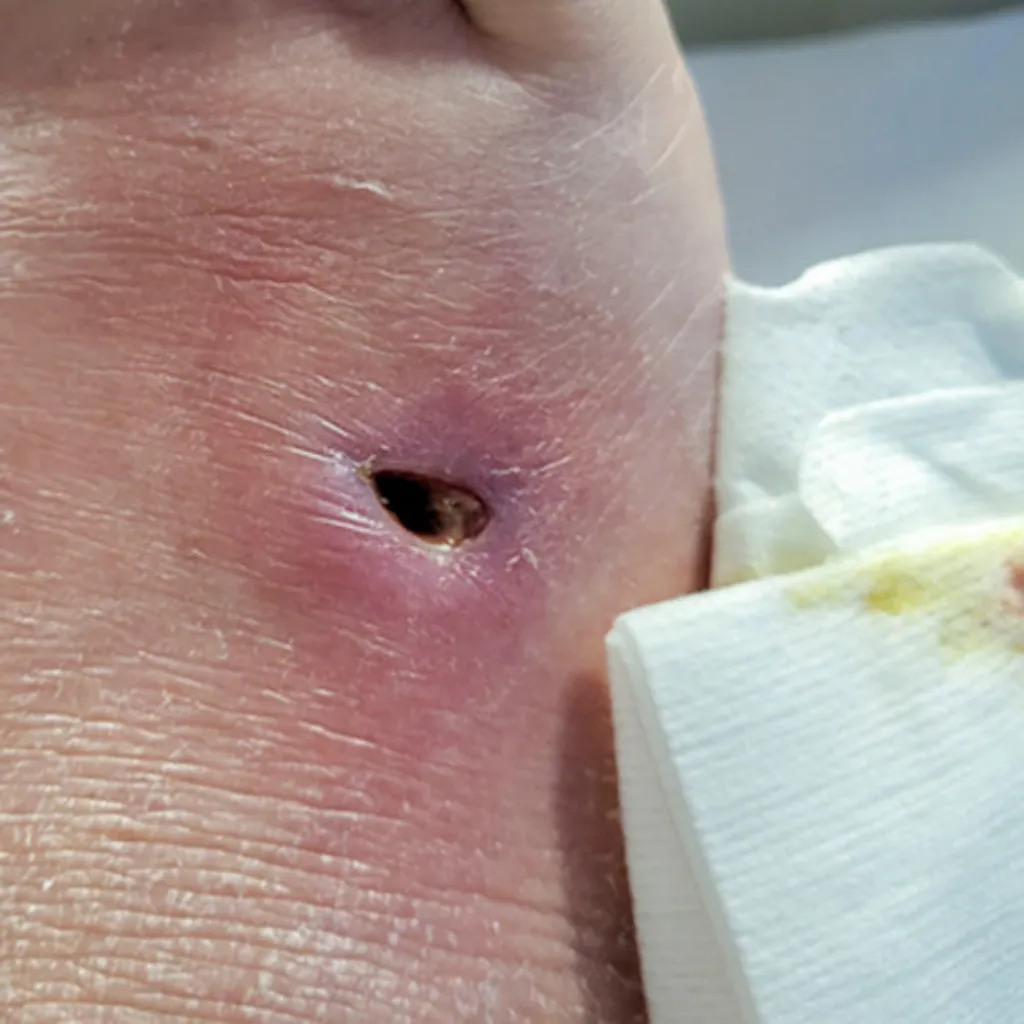
In some cases, fat necrosis may liquefy and form an oil cyst that drains through the skin. Persistent drainage or non-healing wounds require further evaluation.
Differentiating from other conditions
Fat necrosis can resemble an abscess, hematoma, seroma, or malignancy. When findings are atypical or worsening, referral for imaging or surgical evaluation is appropriate.
Patient education
Reassure patients that fat necrosis is benign but emphasize the importance of diagnostic follow-up, especially with breast lesions, to rule out malignancy.
Treatment and management options
Management depends on symptom severity, cosmetic concerns, and diagnostic certainty.
Observation
For asymptomatic, confirmed cases, observation is the most common approach. Periodic clinical exams and imaging may be recommended.
Aspiration
If an oil cyst forms and causes discomfort, needle aspiration may relieve symptoms. Recurrence is possible.
Surgical excision
Surgical removal may be indicated when:
- Diagnosis is uncertain
- The lesion is painful
- There is a significant cosmetic deformity
- The mass persists or enlarges
Management of associated wounds
In cases involving wound breakdown or drainage, wound care principles apply:
- Maintain a clean, moist wound environment
- Monitor for infection
- Use appropriate dressings based on exudate level
- Collaborate with surgical teams as needed
In patients who develop fat necrosis after a BBL or other cosmetic procedure, interdisciplinary coordination is critical to ensure safe and effective recovery.
When to refer
Referral to a surgeon or breast specialist is warranted when:
- Imaging findings are inconclusive
- The mass is enlarging
- There are suspicious skin changes
- The patient has risk factors for malignancy
- Symptoms persist despite conservative management
Timely referral helps differentiate fat necrosis from breast cancer on an ultrasound and ensures appropriate intervention.
By understanding what causes fat necrosis and how it presents, clinicians can provide informed, evidence-based care while reducing patient anxiety and supporting optimal healing outcomes.