Wound Care Treatment for Pilonidal Cysts
Reviewed by: Tara C. Triplett
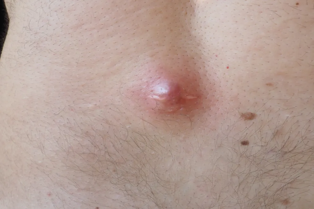
Pilonidal cysts, often occurring in the sacrococcygeal region, present unique challenges in wound management due to their location and tendency for recurrence. As a wound care nurse, understanding the spectrum of treatment options, from conservative management to surgical interventions, is crucial for optimizing patient outcomes. This guide delves into the various treatment modalities, emphasizing the nurse's role in each phase of care.
Conservative management strategies
Conservative treatments may be effective in the early stages or for non-infected pilonidal cysts.
Hair removal
Regular hair removal in the gluteal cleft can prevent hair from penetrating the skin and forming cysts. Methods include shaving, depilatory creams, or laser hair removal. Laser hair removal has shown promise in reducing recurrence rates.
Hygiene and skin care
Maintaining cleanliness in the affected area is vital. Patients should be advised to:
- Clean the area daily with mild soap and water.
- Avoid prolonged sitting to reduce pressure on the area.
- Wear loose-fitting clothing to minimize friction.
- Take measures to control moisture and incontinence.
Incision and drainage (I&D)
For acute abscesses, I&D is the first-line treatment. This procedure involves:
- Making a small incision to drain pus and relieve pressure.
- Cleaning the cavity to remove debris.
Post-procedure, the wound is typically left open to heal by secondary intention. Wound care nurses play a pivotal role in:
- Educating patients on wound care techniques.
- Monitoring for signs of infection or complications.
- Ensuring proper dressing changes to promote healing.
Surgical interventions
Surgical options are considered when conservative measures fail, with the presence of a complex abscess and sinus tracts, or in cases of recurrent disease.
Excision with primary closure
This involves removing the cyst and closing the wound with sutures. While this method offers faster healing, it carries a higher risk of recurrence.
Excision with secondary intention healing
The wound is left open post-excision, allowing it to heal naturally. This method reduces recurrence rates but requires diligent wound care over an extended period.
Flap procedures
There are several flap procedures and advanced techniques a surgeon may use to manage complex or recurrent pilonidal cysts. Below are two examples:
Limberg flap
The Limberg flap involves the excision of the pilonidal sinus in a rhomboid (diamond) shape, followed by rotation of a nearby flap of skin and subcutaneous tissue to cover the defect. The goals are to:
- Flatten the natal cleft, which reduces hair accumulation and friction.
- Improve wound healing by bringing in healthy tissue with good blood supply.
- Minimize tension on the surgical site, which supports a more stable closure.
Wound care nurses should monitor for:
- Flap viability (check for signs of necrosis, poor perfusion, or excessive tension).
- Seroma or hematoma formation under the flap.
- Infection or dehiscence at the suture line.
Karydakis flap
The Karydakis technique uses elliptical excision to remove the cyst and sinus tracts, then mobilizes a lateral flap of tissue to cover the midline defect. Unlike the Limberg flap, this method displaces the surgical closure off the midline, which is particularly helpful since midline closures have higher failure and recurrence rates.
Benefits of the Karydakis flap include:
- Faster healing time compared to open excision.
- Reduced postoperative discomfort.
- Lower recurrence rate due to the off-midline closure.
Postoperative care considerations for nurses include:
- Ensuring the patient avoids pressure on the surgical site for several weeks.
- Monitoring for fluid collection and providing appropriate drainage if necessary.
- Supporting the patient's gradual return to activity to avoid wound disruption.
Both flap techniques have high success rates when appropriately managed. However, due to their complexity, these procedures demand a higher level of post-operative wound care vigilance from nursing staff.
Preventing recurrence: A top priority in long-term care
Preventing recurrence is one of the most important responsibilities for wound care nurses managing patients with pilonidal disease. Recurrence rates for pilonidal cysts can be high, ranging from 10% to 30% depending on treatment method and patient adherence to care protocols.
Here’s how wound care nurses can take proactive steps to reduce recurrence risk:
1. Consistent hair management
- Shaving or depilatory creams: Teach patients to keep the area free of hair regularly, but caution against shaving too aggressively, which can cause microtrauma.
- Laser hair removal: Consider recommending laser therapy for patients with dense or coarse hair, especially those with a history of recurrence. Multiple studies have shown that laser removal significantly lowers recurrence rates.
2. Skin care and hygiene
- Encourage patients to shower daily and clean the sacrococcygeal area with a gentle, non-irritating cleanser.
- Ensure thorough drying after bathing to avoid skin maceration.
- Apply a barrier cream if patients are prone to sweating or moisture build-up.
3. Pressure management
- Suggest using cushions or sitting on soft surfaces to relieve pressure on the healing area during the recovery period.
- Educate patients who work in seated positions (e.g., drivers, desk workers) about taking frequent breaks to stand and stretch.
4. Clothing and environment
- Advise against tight-fitting clothing that may cause friction or trap sweat in the gluteal cleft.
- Recommend breathable fabrics like cotton to reduce moisture accumulation.
5. Weight management
- Discuss the link between obesity and pilonidal disease. Excess adipose tissue can deepen the gluteal cleft and increase friction and hair entrapment.
- Offer referrals to nutrition services or lifestyle programs if appropriate.
6. Early symptom reporting
- Encourage patients to report any signs of tenderness, swelling, or drainage early.
- Provide education on self-assessment techniques so that they can identify symptoms before a full recurrence occurs.
7. Follow-up and surveillance
- Schedule regular follow-ups for patients’ post-treatment, especially after surgical intervention.
- Document changes in wound appearance, drainage, and healing progress thoroughly.
- Work collaboratively with primary care providers or surgical teams for prompt intervention if signs of recurrence emerge.
References
- Cork Medical. Wound Care Options for the Pilonidal Cyst. https://www.corkmedical.com/post/wound-care-options-for-the-pilonidal-cyst
- WoundSource. Pilonidal Cysts: Pathophysiology, Wound Care Management, and Patient Education. https://www.woundsource.com/blog/what-you-need-know-about-pilonidal-cysts
- American Family Physician. Pilonidal Disease Management: Guidelines from the ASCRS. https://www.aafp.org/pubs/afp/issues/2019/1101/p582.html
- Pilonidal Support Alliance. Wound Dressing. https://www.pilonidal.org/surgery-aftercare/wound-dressing/
- Mayo Clinic. Pilonidal cyst. https://www.mayoclinic.org/diseases-conditions/pilonidal-cyst/diagnosis-treatment/drc-20376332